Fasciculus:Plasmacytoma ultramini1.jpg
Mensura huius perspectionis: 800 × 589 elementa imaginalia. Aliae mensurae: 320 × 236 elementa imaginalia | 640 × 472 elementa imaginalia | 950 × 700 elementa imaginalia.
Sua resolutio (950 × 700 elementa imaginalia, magnitudo fasciculi: 360 chiliocteti, typus MIME: image/jpeg)
Summarium
| Descriptio |
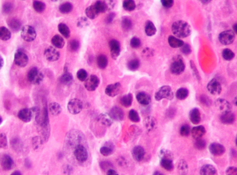
English: Micrograph of a plasmacytoma. H&E stain.
The micrograph shows abundant (malignant) plasma cells with the occasional Mott cell, a plasma cell with intracytoplasmic Russell bodies (an eosinophilic uniformly staining membrane bound body which contains immunoglobulin). Other features of plasmacytomas (not apparent on the image) are:
Multiple myeloma (which is diagnosed using several clinical criteria) is, histologically, a plasmacytoma. Related images
|
| Fons | Opus proprium |
| Auctor | Nephron |


Potestas usoris
I, the copyright holder of this work, hereby publish it under the following licenses:
This file is licensed under the Creative Commons Attribution-Share Alike 3.0 Unported license.
- Tibi licet:
- communicare – copiare, distribuere et committere hoc opus
- to remix – to adapt the work
- His condicionibus:
- attributio – You must give appropriate credit, provide a link to the license, and indicate if changes were made. You may do so in any reasonable manner, but not in any way that suggests the licensor endorses you or your use.
- aequa parte – If you remix, transform, or build upon the material, you must distribute your contributions under the same or compatible license as the original.

|
Licet hoc documentum exscribere vel distribuere vel demutare sub GNU Liberarum Litterarum Licentiae conditionibus in editione 1.2 aut in ulla editione recentiori a Fundatione Liberarum Programmationis Partium publicata; praeterquam Sectiones Immutabiles et Verba Involucra Adversa et Aversa. Licentiae exemplar praesto est in sectione intitulata GNU Free Documentation License. |
Tibi typum permissionis ligere licet.
Historia fasciculi
Presso die vel tempore fasciculum videbis, sicut tunc temporis apparuit.
| Dies/Tempus | Minutio | Dimensiones | Usor | Sententia | |
|---|---|---|---|---|---|
| recentissima | 03:34, 21 Iulii 2009 | | 950 × 700 (360 chiliocteti) | Nephron | {{Information |Description={{en|1=Micrograph of a '''plasmacytoma'''. H&E stain. The micrograph shows abundant (malignant) plasma cells with the occasional ''Mott cell'', a plasma cell |
Nexus ad fasciculum
Ad hunc fasciculum nectunt:
Usus fasciculi per inceptus Vicimediorum
Quae incepta Vici fasciculo utuntur:
- Usus in ar.wikipedia.org
- Usus in azb.wikipedia.org
- Usus in bs.wikipedia.org
- Usus in ca.wikipedia.org
- Usus in cs.wikipedia.org
- Usus in el.wikipedia.org
- Usus in en.wikipedia.org
- Usus in en.wiktionary.org
- Usus in es.wikipedia.org
- Usus in fa.wikipedia.org
- Usus in fr.wikipedia.org
- Usus in ga.wikipedia.org
- Usus in gl.wikipedia.org
- Usus in ha.wikipedia.org
- Usus in he.wikipedia.org
- Usus in hi.wikipedia.org
- Usus in hu.wikipedia.org
- Usus in ia.wikipedia.org
- Usus in it.wikipedia.org
- Usus in ko.wikipedia.org
- Usus in ms.wikipedia.org
- Usus in nl.wikipedia.org
- Usus in or.wikipedia.org
- Usus in pt.wikipedia.org
- Usus in ru.wikipedia.org
- Usus in simple.wikipedia.org
- Usus in sl.wikipedia.org
- Usus in sr.wikipedia.org
- Usus in sv.wikipedia.org
- Usus in ta.wikipedia.org
View more global usage of this file.