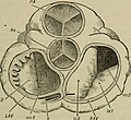
Fasciculus:Orifices of the Heart seen from above in An academic physiology and hygiene (1903).jpg

Sua resolutio(1 612 × 1 472 elementa imaginalia, magnitudo fasciculi: 414 chiliocteti, typus MIME: image/jpeg)

Hic fasciculus apud Vicimedia Communia iacet; in aliis inceptis adhiberi potest. Contenta paginae descriptionis fasciculi subter monstrantur.

Summarium

Descriptio
English:

Identifier: academicphysio00bran (find matches)
Title: An academic physiology and hygiene ..
Year: 1903 (1900s)
Authors: Brands, Orestes M. (from old catalog) Van Gieson, Henry C., (from old catalog) joint author
Subjects: Hygiene Physiology
Publisher: Boston, B. H. Sanborn & co
Contributing Library: The Library of Congress
Digitizing Sponsor: The Library of Congress

View Book Page: Book Viewer
About This Book: Catalog Entry
View All Images: All Images From Book
Click here to view book online to see this illustration in context in a browseable online version of this book.

Text Appearing Before Image:
Fig. 31. Interior of the Right Side of the Human Heart. EXPLANATION. i, superior vena cava ; 2, inferior vena cava ; 3, interior of the right auricle ; 4, semi-lunar valves of the pulmonary artery ; a?, papillary muscle ; 5, 5, and 5, cusps of the tri-cuspid valve ; 6,pulmonary artery ; 7, 8, and 9, the aorta and its branches ; 10, left auricle;11, left ventricle. is especially true of the left ventricle, whose work it is topropel the blood through the whole system. THE BLOOD AND ITS CIRCULATION. 85 10. Besides the valves already mentioned, there are also valves in the aorta (the main trunk of the arterial system),and in the pulmonary artery (the artery conveying bloodto the lungs). nw2
Text Appearing After Image:
Iu3 RAV Fig. 32. The Orifices of the Heart seen from above, the Auricles and Great Vesselsbeing cut away. EXPLANATION. P. A, pulmonary artery, and Ao, aorta, with their semilunar valves. R. A. !r., right ■ do-ventricular orifice, showing the three folds (/v, 1, 2, 3) of the tricuspid valve. L. A. I., left auriculo-vcntriadar orijice, showing its mitral valve of IWO flaps at ;//. v., 1 and 2. A piece of whalebone at b passes into the coronary vein. The tooth-like appeafiUM * the left side of L. A. I, is due to the fact that the section of the auricle is carried through -een on the exterior surface of the heart. 11. In a healthy condition of the heart, all of thesevalves work harmoniously and perfectly; but in certain _ mic dis they become deranged, thus leading to and often fatal results. 12. Course of the Blood in Circulation. — The minute veinsin all parts of the body collect the impure blood. Thveinlets unite and form larger branches which finally oil- 86 ACADEMIC PHYSIOLOGY. mi

Note About Images

Please note that these images are extracted from scanned page images that may have been digitally enhanced for readability - coloration and appearance of these illustrations may not perfectly resemble the original work.
Datum
Fons

https://www.flickr.com/photos/internetarchivebookimages/14594429420/

Auctor Internet Archive Book Images
Permissio
(Reusing this file)
At the time of upload, the image license was automatically confirmed using the Flickr API. For more information see Flickr API detail.
Flickr tags
InfoField
Flickr posted date
InfoField
30 Iulius 2014

Potestas usoris

This image was taken from Flickr's The Commons. The uploading organization may have various reasons for determining that no known copyright restrictions exist, such as:
  1. The copyright is in the public domain because it has expired;
  2. The copyright was injected into the public domain for other reasons, such as failure to adhere to required formalities or conditions;
  3. The institution owns the copyright but is not interested in exercising control; or
  4. The institution has legal rights sufficient to authorize others to use the work without restrictions.

More information can be found at https://flickr.com/commons/usage/.


Please add additional copyright tags to this image if more specific information about copyright status can be determined. See Commons:Licensing for more information.
This image was originally posted to Flickr by Internet Archive Book Images at https://flickr.com/photos/126377022@N07/14594429420. It was reviewed on 18 September 2015 by FlickreviewR and was confirmed to be licensed under the terms of the No known copyright restrictions.

18 September 2015

Captions

Add a one-line explanation of what this file represents

Items portrayed in this file

depicts Anglica

MIME type Anglica

image/jpeg

Historia fasciculi

Presso die vel tempore fasciculum videbis, sicut tunc temporis apparuit.

Dies/TempusMinutioDimensionesUsorSententia
recentissima10:07, 18 Septembris 2015Minutum speculum redactionis 10:07, 18 Septembris 2015 factae1 612 × 1 472 (414 chiliocteti)== {{int:filedesc}} == {{subst:chc}} {{information |description={{en|1=<br> '''Identifier''': academicphysio00bran ([https://commons.wikimedia.org/w/index.php?title=Special%3ASearch&profile=default&fulltext=Search&search=insource%3A%2Facademicphysio00b...

Usus fasciculi per inceptus Vicimediorum

Quae incepta Vici fasciculo utuntur: